Tear Drainage System (Lacrimal System) & Treatment
Anatomy:
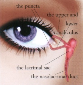
The tear drain consists of two small openings called the puncta, one in your upper and the other in your lower eyelid. Each of these openings leads into a small tube called a canaliculus, which, in turn, empties into the tear sac at the inside corner of your eye along your nose. An opening into the lacrimal sac leads into a canal called the nasolacrimal duct, which passes through the bony structures surrounding your nose and empties tears into your nasal cavity (see Figure 7).
How does the tear drain work?
The tears pass through the lacrimal drainage system by blinking and the principle of capillary action. When blinking, your lids push tears evenly across your eyes to keep your eyes moist and healthy. Blinking also pumps tears into the puncta, and by capillary action, they are drawn into the lacrimal sac, which travels through the tear duct and drains into your nose. If the tear duct is blocked or the capillary action of the tears is not working, your tears back up and spill over your eyelids as if you are crying. Tears trapped in the tear sac also become stagnant and infected like a pond in the forest when the water has sat there for a period of time.
What are the symptoms of having a plugged tear drain?
Many times the symptoms revolve around an infected tear sac and duct. The most common symptoms of this are excessive watering, mucous discharge, eye irritation, and a painful swelling in the inner corner of your eyelid. If the symptoms go untreated, infection can develop around the entire area of your eye.
What is the treatment?
We may recommend any number of treatments based on the analysis of your symptoms and your diagnostic tests. In some instances, it may be as simple as applying warm compresses with topical and oral antibiotics, but often surgery is the most effective treatment. If the obstruction is partial in nature, or involves a poor pump mechanism, sometimes endoscopic nasal surgery with placement of Crawford tubes can be done. This is a surgery that is done in the hospital, but has a very minimal recovery time. This is reserved only for smaller obstructions and patients with lax eyelids with a poor pump. Sometimes an eyelid tightening procedure is needed in conjunction with the placement of tubes or in and of itself. Tightening the eyelids helps the advancement of tears through the tear drainage system by strengthening the pump mechanism.
Larger (complete) obstructions are more common, and the surgical solution is dacryocystorhinostomy (DCR). The DCR has been around for over 100 years and has a success rate of over 90% for adults who have had no prior nasal surgery or disease. The surgery is an outpatient procedure and is usually performed after the inflammation is reduced. This is done in the hospital under general anesthesia, and your recovery at home generally is in about a week. To perform the DCR, we will create a new tear drain opening from the lacrimal sac directly into your nose that allows tears to bypass the obstruction. An incision is usually made from the skin surface, and sometimes from just inside the nose
A silicone tube may be placed temporarily (usually six to twelve months) in the new tear drain to keep it open while the healing process occurs. Rarely the obstruction is beyond repair and in this instance it is necessary to surgically place a tiny artificial drain called a Jones tube behind the inner corner of the eyelid. This tube is made of glass and remains permanently in the tear duct.
What are the risks and complications?
Risks and complications are associated with the usual risks of anesthesia and surgery including bleeding and infection. Occasionally, the body may form scar tissue, which blocks the drain again, which may require repeating the procedure.